Kinesiology
|
Anatomy
Bones provide attachment points and support for muscles. Bones are connected together by fibrous tissue called Ligaments. Tendons are also fibrous tissue and attach muscle to bone. Both have some elasticity and do not heal on their own if torn. An overstretched tendon is called tendonitis and is an inflamed tendon. Cartilage is also fibrous tissue but is not elastic. Cartilage is used to cushion the junction of two bones.
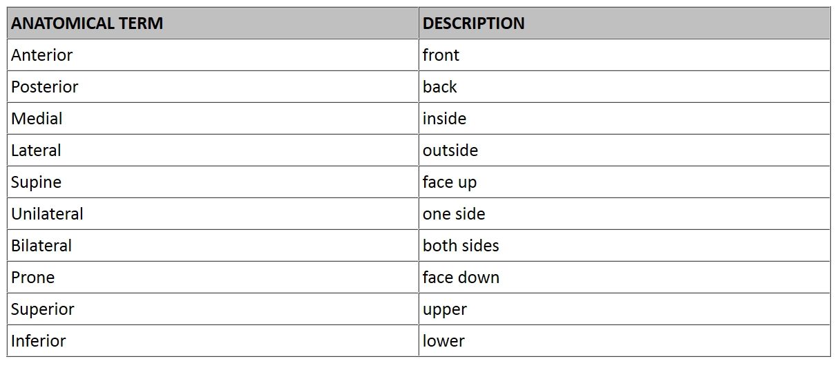
The body is divided into three anatomical planes the Frontal, Sagittal and Horizontal. The Frontal plane divides the body from front to back. The Sagittal plane divides the body down the center or vertically. The Horizontal plane divides upper and lower. The table below lists the anatomical term and the corresponding description.
|
Muscle Action
The three types of muscle contraction are Isometric, Isotonic, and Isokinetic. Isometric is defined as that type of contraction where muscle tension and muscle length remain constant. This type of exercise provides muscle strength gains but only at the joint angle held during the exercise. Isotonic contraction is defined as that where the muscle tension remains constant and muscle length varies. Isokinetic contraction is defined as varying tension and length.
In each exercise there are four main functions of the associated muscles, Agonists (prime movers), Antagonists, Stabilizers and Assistors. The Agonists is generally the muscle we are exercising. The Antagonist is the opposing muscle and acts in contrast to the agonist. The Stabilizer muscles are those that hold a joint in place so that the exercise may be performed. The Assistors help the Agonist muscle doing the work. The stabilizer muscles are not necessarily moving during exercise, but provide stationary support.
For example, when doing biceps curls, the biceps are the agonists, the triceps are the antagonists and various muscles including the deltoids are the stabilizer muscles. However, when doing a triceps push down, now the triceps are the agonists and the biceps are the antagonists. Again the deltoid muscles are the stabilizer muscles. The agonist/antagonist relationship changes depending on which muscle is expected to do the work. However, every muscle group has an opposing muscle group. The following table lists muscles and their opposing counterparts:
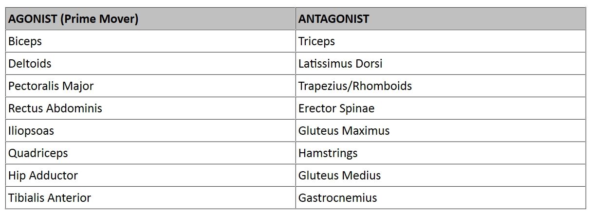
In reference to Agonist and Antagonist, this above list could easily be reversed when exercising the muscles in the right hand column. Muscle balance is that relationship between the Agonist and Antagonist. It is important to have muscle balance to prevent injury. If the Agonist is much stronger than the Antagonist, the Agonist can overpower and injure the Antagonist.
Tendons are made up of fibrous tissue and connect muscle to bone. Tendonitis is an inflammation of the tendon due to overuse. A stretching or tearing of the tendon is referred to as a strain. A Strain is a muscle or tendon injury.
Ligaments are also fibrous tissue and connect bone to bone. There are less flexible than tendons. The function of ligaments is to restrict the joint movement within normal parameters. When a ligament is over stretched or torn it is called a sprain. Since ligaments don't have a vascular system, they may take a very long time to repair or may never return to their original length. This can cause abnormal joint movement and even cartilage and bone wear due to this unrestricted movement.
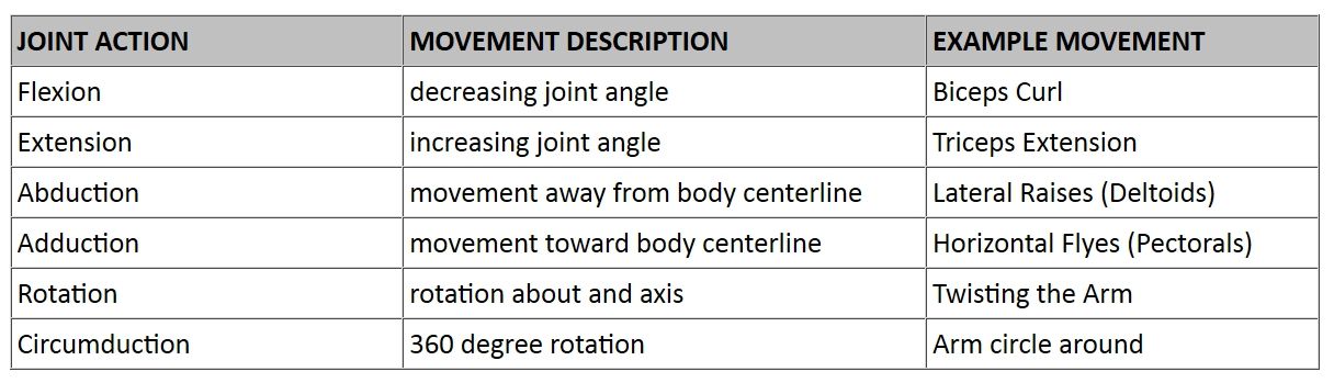
Joint Action
Joints provides a fulcrum point for muscles to do work. There are six types of joint action:
|